Images and licences
The diagrams in the Knowledge articles are drawn by FFmove and have no other author.
The other images belong to the authors below and are used only when they are in the public domain or under the Creative Commons CC BY or CC BY-SA licences. Each image lists its author, its licence, the page of the source file, the changes made by FFmove and the pages where it appears. An image adapted from a CC BY-SA file stays under CC BY-SA.
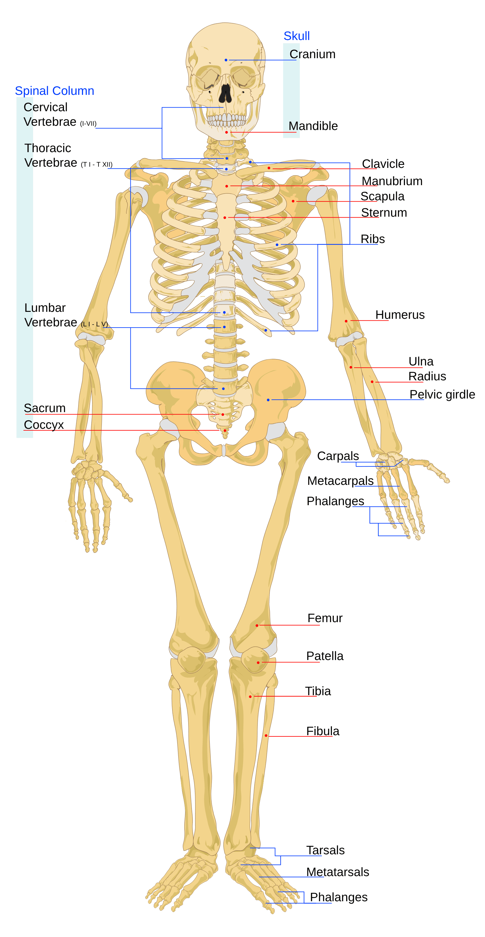
The human skeleton, anterior view
- Author
- LadyofHats Mariana Ruiz Villarreal
- Licence
- Public domain
- Source
- File:Human skeleton front en.svg · Wikimedia Commons
- Changes
- converted to another format, resized
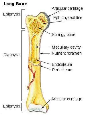
The structure of a long bone
- Author
- NCI SEER Training Modules (training.seer.cancer.gov)
- Licence
- Public domain
- Source
- File:Illu long bone.jpg · Wikimedia Commons
- Changes
- converted to another format

The knee joint
- Author
- Henry Vandyke Carter
- Licence
- Public domain
- Source
- File:Gray345.png · Wikimedia Commons
- Changes
- none
- Appears in
- Classification of joints

Diagram of a synovial joint
- Author
- Madhero88
- Licence
- CC BY-SA 3.0
- Source
- File:Joint.svg · Wikimedia Commons
- Changes
- converted to another format, resized
- Appears in
- Classification of joints
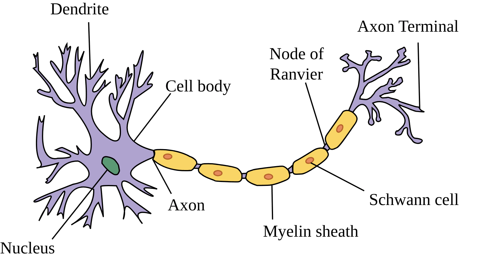
The neuron
- Author
- User:Dhp1080
- Licence
- CC BY-SA 3.0
- Source
- File:Neuron.svg · Wikimedia Commons
- Changes
- converted to another format, resized
- Appears in
- Nervous control of movement

Superficial muscles, anterior view
- Author
- Mikael Häggström
- Licence
- Public domain
- Source
- File:Muscles anterior labeled.png · Wikimedia Commons
- Changes
- resized
- Appears in
- Trunk and shoulder-girdle stability

Superficial muscles, posterior view
- Author
- Mikael Häggström
- Licence
- Public domain
- Source
- File:Muscle posterior labeled.png · Wikimedia Commons
- Changes
- none
- Appears in
- Trunk and shoulder-girdle stability
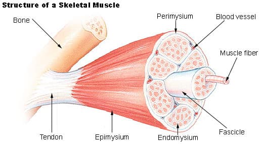
The structure of skeletal muscle
- Author
- NCI SEER Training Modules (training.seer.cancer.gov)
- Licence
- Public domain
- Source
- File:Illu muscle structure.jpg · Wikimedia Commons
- Changes
- converted to another format
- Appears in
- Muscle architecture
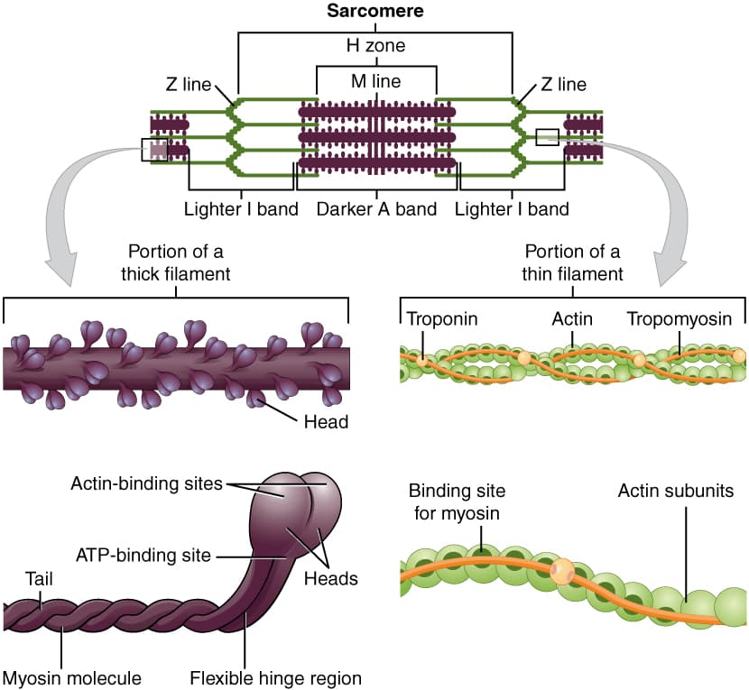
The thick and thin filaments of the sarcomere
- Author
- OpenStax
- Licence
- CC BY 4.0
- Source
- File:1003 Thick and Thin Filaments.jpg · Wikimedia Commons
- Changes
- converted to another format
- Appears in
- Muscle architecture
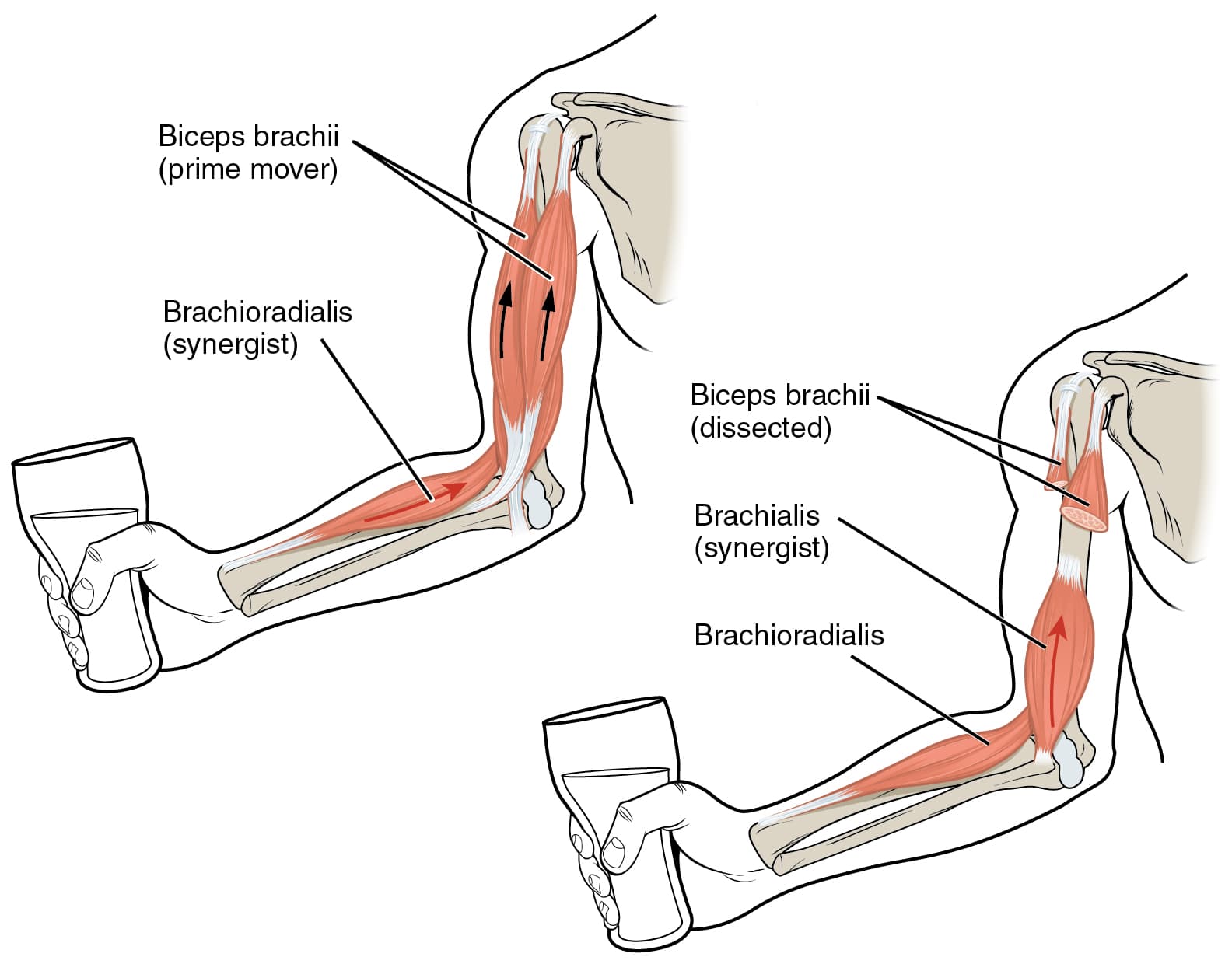
The prime mover and the synergists in elbow flexion
- Author
- OpenStax College
- Licence
- CC BY 4.0
- Source
- File:Biceps Muscle CNX.jpg · Wikimedia Commons
- Changes
- converted to another format
- Appears in
- Principles of functional training
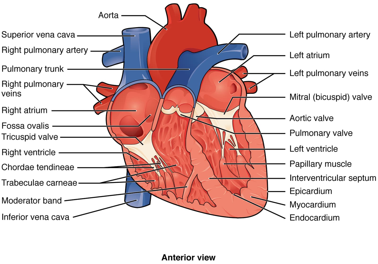
The internal anatomy of the heart
- Author
- OpenStax College
- Licence
- CC BY 3.0
- Source
- File:2008 Internal Anatomy of the HeartN.jpg · Wikimedia Commons
- Changes
- resized, converted to another format
- Appears in
- The heart and circulation

The events of the cardiac cycle
- Author
- adh30 revised work by DanielChangMD who revised original work of DestinyQx; Redrawn as SVG by xavax
- Licence
- CC BY-SA 4.0
- Source
- File:Wiggers Diagram 2.svg · Wikimedia Commons
- Changes
- converted to another format, resized
- Appears in
- The heart and circulation

The circulatory system
- Author
- LadyofHats, Mariana Ruiz Villarreal
- Licence
- Public domain
- Source
- File:Circulatory System en.svg · Wikimedia Commons
- Changes
- converted to another format, resized
- Appears in
- The heart and circulation
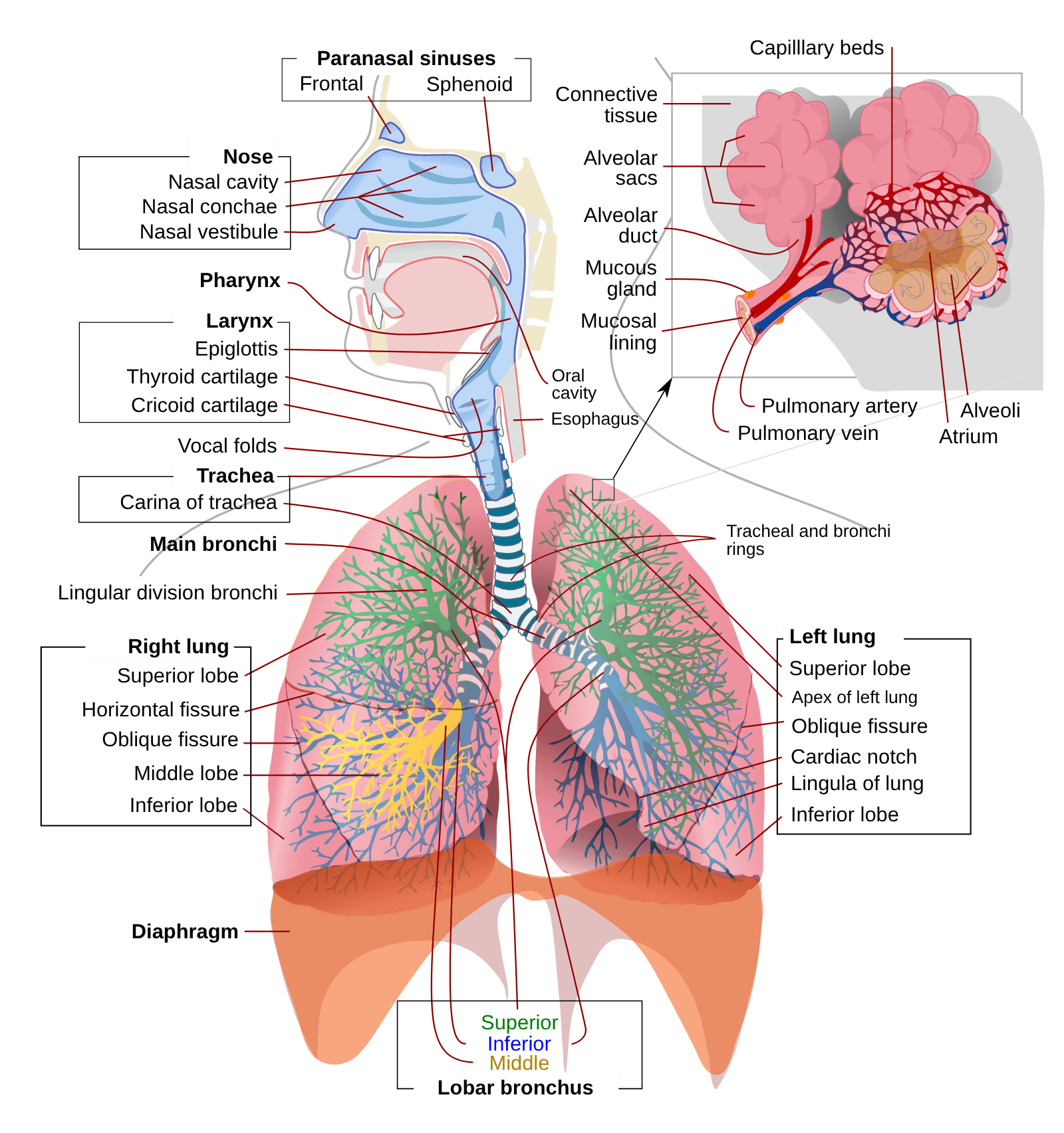
The respiratory system
- Author
- LadyofHats, Jmarchn
- Licence
- Public domain
- Source
- File:Respiratory system complete en.svg · Wikimedia Commons
- Changes
- converted to another format, resized
- Appears in
- Mechanics of breathing
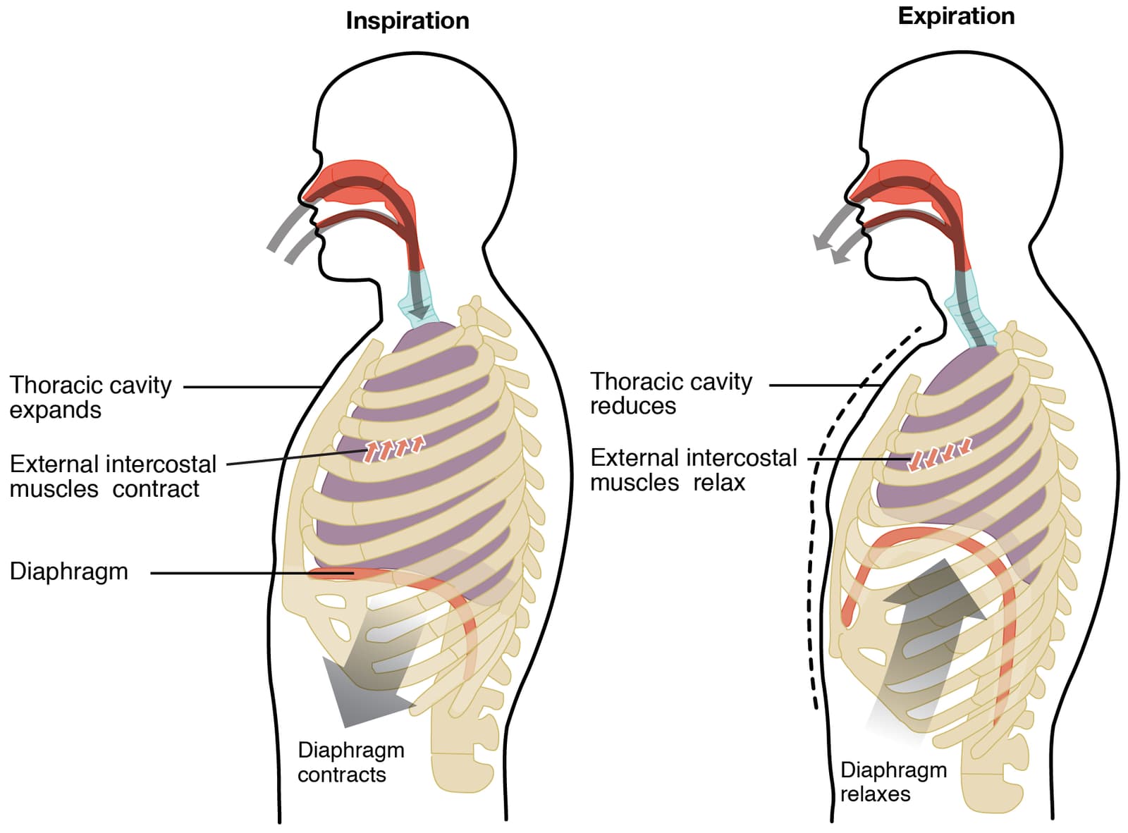
Inspiration and expiration
- Author
- OpenStax College
- Licence
- CC BY 3.0
- Source
- File:2316 Inspiration and Expiration.jpg · Wikimedia Commons
- Changes
- resized, converted to another format
- Appears in
- Mechanics of breathing